
The 3rd International Nursing and Health Sciences Students and Health Care Professionals Conference (INHSP)
More infoTable salt (sodium chloride) is an ionic compound consisting of positive ions (cations) and negative ions (anions) to form neutral compounds that can provide a healing effect on wounds. This study's purpose of seeing and test the impact of soaking 7% sodium chloride concentration on people's salt toward the wound healing process.
MethodThis study was an experimental laboratory using the One-Way ANOVA test and the Mann Whitney test conducted in the animal enclosure of the Faculty of Pharmacy, Hasanuddin University, Makassar. The study was conducted from July to August 2019. Samples of 20 mice (Mus Musculus) female swiss webster strains were sliced on the abdominal skin then divided into two groups: the treatment group (n=15) and the control group (n=5). The wound area was observed from the first day to the seventh day to see the wound closure process.
ResultsThe research shows that soaking 7% of table salt concentration can significantly accelerate the wound healing process compared to the control group, with a decrease in wound diameter on the 3rd day and completely heal on the 7th day.
Conclusion7% concentration of table Salt Soaking can increase the effectiveness of wound healing.
The wound healing process is a quality of tissue life process associated with the regeneration of tissue. The physiological process takes place immediately after the wound occurs by activating the blood of the coagulation system. The release of mediators from platelets, namely Platelet Activating Factor (PAF) and Thromboxane which are then attached to the wound tissue that converts fibrin to fibrinogen so that blood clots last 5–10min and after that vasodilation will occur, capillary nerve stimulation synthesis (local sensory-nerve endings) occur in the inflammatory phase which ends on the 3rd day to 4th day.
In the phase of tissue structure, improvement marked by angiogenesis occurs in the proliferation or re-epithelialization phase from 3rd day or 4th day and maximum to 14th day. The final phase of wound healing occurs in 21 days to 1 year where there is a balance between the collagen produced and the one that is broken.1,2 The wound healing process normally occurs if supported by nutrition, immune system, and wound care. Wound care using natural materials has been done a long time ago, including wound care using sodium chloride from seawater. Soaking wounds in 7% table salt concentration with osmotic salt properties can provide a drying effect on the wound so that the growth of new tissue accelerates skin contact more quickly. Bacteriostatic salt properties in which streptococcus aureus causes the infection is unable to survive on sodium chloride, which has a concentration of 7%.3
The 7% table salt concentration in this study was made in a liquid to wash the wound, which provides wound drying and anti-bacterial properties, so the wound healing process becomes faster. This study wanted to identify the effect of a 7% concentration of table salt immersion on wound healing in the stomach of female mice (Mus Musculus).
MethodThe 7% table salt concentration is made from people's salt. It is produced by salt farmers in Maccini Baji village, Labbakkang sub-district, Pangkep district, and then makes the salt concentration in the oceanography laboratory of the Faculty of Oceanography Hasanuddin University. A dose of 70g of salt dissolved in boiled water into a 1-l sodium chloride solution. Furthermore, this concentration is used to soak the incision wound in mice with an incision area of approximately 1.5cm with a depth of all layers of skin and muscle cut off then sewn using catgut, soaking process done once every day for seven days. Observation and measurement are carried out until the wound heals.
The research subjects were Swiss Webster female mice aged 8–10 weeks with a weight of 20–25g, totaling twenty animals from the Laboratory of Animal Medicine Faculty of Hasanuddin University, by considering the cleanliness of the cage, the area of the cage by placing 5 mice for each group and adapted for one week, with enough food and drink every day. The mice were anesthetized by using a ketamine injection with a dose of 0.2ml/kg BW before an abdominal wound was performed. After being anesthetized, the subject's abdominal surface is smeared with veet then the hair is shaved and cleaned with 70% alcohol. The cut was made by drawing using a ruler and a marker with a length of 1.5cm. Then after mice are unconscious, an incision is made in the stomach of the mice with the help of Chirurgie nippers, a scalpel, and scissors to made a wound on the part that has been drawn. The skin and subcutaneous tissue are removed using tweezers until the mole's intestine is visible, then sewn back using a catgut with 1–3 stitches.
Mice were divided into two groups: the control and treatment groups that were given a 7% concentration of table salt soaking. The Control group was divided into three groups – each group consisted of 5 mice. Then the treatment group was immersed for 5min, 10min, and 15min at noon every day. From the observation result, the wound area on the 1st day and second day has not changed. On the 3rd day, the wound began to appear in the process of drying the wound, and the diameter of the wound was getting smaller and mostly healed on the 7th day. Measuring the diameter of the wound is done every day, starting from the 1st day to 7th day.
Data analysis was performed using a one-way ANOVA test to determine whether there were differences in the average area of cut wounds in the control and treatment groups. Then an independent t-test is performed if the data is normally distributed or the Mann–Whitney test if the data is not normally distributed. Finally, the data normality test uses the Shapiro–Wilk test. The significance of the test results is determined based on a p-value <0.05.
This research has been tested and deemed feasible to be carried out by the Health Research Ethics Commission of the Medical Faculty of Hasanuddin University through letter Number: 541/UN4.6.4.5.31/PP36/2019.
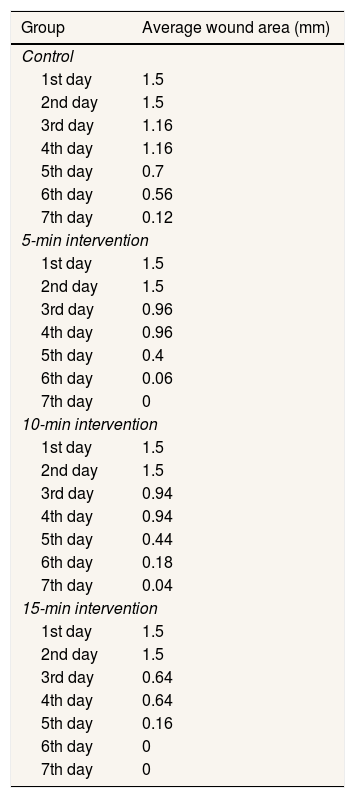
ResultsTo find out the depiction of the results of observations that have been obtained, then carried out a descriptive analysis of the area of the wound in mice from each control group and treatment group, namely intervention 5min, 10min, and 15min. The following table compares the area of wounds between the control group and the treatment group.
Comparing the average injuries between the control group and the treatment group began to be different on the 3rd day to the 7th day. The descriptive analysis shows that the wound in the treatment or intervention group is faster closed than the area of the wound in the control group (Table 1).
Comparison of wound areas in the control and treatment groups.
| Group | Average wound area (mm) |
|---|---|
| Control | |
| 1st day | 1.5 |
| 2nd day | 1.5 |
| 3rd day | 1.16 |
| 4th day | 1.16 |
| 5th day | 0.7 |
| 6th day | 0.56 |
| 7th day | 0.12 |
| 5-min intervention | |
| 1st day | 1.5 |
| 2nd day | 1.5 |
| 3rd day | 0.96 |
| 4th day | 0.96 |
| 5th day | 0.4 |
| 6th day | 0.06 |
| 7th day | 0 |
| 10-min intervention | |
| 1st day | 1.5 |
| 2nd day | 1.5 |
| 3rd day | 0.94 |
| 4th day | 0.94 |
| 5th day | 0.44 |
| 6th day | 0.18 |
| 7th day | 0.04 |
| 15-min intervention | |
| 1st day | 1.5 |
| 2nd day | 1.5 |
| 3rd day | 0.64 |
| 4th day | 0.64 |
| 5th day | 0.16 |
| 6th day | 0 |
| 7th day | 0 |
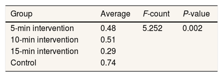
The differences in the average area of cut wounds in the treatment and control groups, a one-way ANOVA analysis was performed. The analysis was performed on the area of the wound area on the 3rd to 7th day for each treatment and control group. This is because there is no change in the field of the wound on days one and two, so it does not provide meaningful information. In addition, if the recording of days one and two are included in the analysis, it can obscure the results of the analysis of variance that is carried out. The results are as follows (Table 2).
Based on Table 2, it is known that the average wound area for the 5-minute intervention group is 0.48, the 10-min intervention is 0.51, the 15-min intervention is 0.29, and the control group is 0.74. From the results of the average wound area in the intervention or treatment group, it is known that the longer the intervention is carried out, the average wound area is also getting smaller. In addition, the average size of the wound area that has been given intervention or treatment is also smaller than the average wound area in the control group. From the results of one-way ANOVA analysis, the F-count is 5.252, and the P-value is 0.002. Because the P-value is smaller than (P<0.05), it can be concluded that there is a significant difference in the average area of mice wounds in the treatment group and the control group.
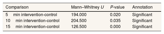
The Mann–Whitney test was performed to determine the significance of the difference in the average wound area of each treatment group to the control group if the data did not have a normal distribution. The analysis was performed on the area of the wound area on the 3rd to 7th day for each treatment and control group. The results are as follows.
Based on Table 3, it is known that the P-value from the Mann–Whitney test results for the comparison of the 5-min intervention group and the control group is 0.020, the 10-min intervention group and the control group is 0.035, and the 15-min intervention group and the control group are 0.000. Because of the P-value of (P<0.05), it can be concluded that the average wound area in each intervention group had a significant difference from the control group.
DiscussionThe wound healing process is a complex process that includes a series of biochemical, cellular, and physiological processes. The process goes through several phases, namely the hemostatic, inflammatory, proliferation, and remodeling phases. Proper wound care is one factor in the formation of new tissue. Wound management using natural ingredients as medicine has been widely carried out today, one of which is using seawater.2,4,5
Seawater contains salt, which is composed of sodium atoms and chloride atoms. Types of bonds in the form of sodium chloride compounds are bonds of positive ions and negative ions. In the process of fermentation of food ingredients, sodium chloride is useful to limit the growth of spoilage organisms and prevent the growth of some microorganisms. However, certain bacteria can still grow in high saline solutions. Seawater can be used in the treatment of external wounds, which have an osmotic effect on the tissue so that the wound becomes dry.3,6,7
Some principles in wound healing are that the body's ability to deal with tissue trauma is influenced by the extent of damage and the general condition of each person. The body's response to the wound is more effective if the nutrients are maintained, the body's response to the system to the trauma of blood flow to the wound tissue, the integrity of the skin and mucous membranes are prepared as the first line to defend against microorganisms, wound healing is enhanced when the wound is free of foreign matter including bacteria.8–10
In this study, wound healing significantly increased in the treatment group compared to the control group (P<0.05). In the control and treatment groups, there were differences in the extent of the wound on the 3rd day to the 7th day. From the results of the one-way ANOVA analysis, the P-value is 0.002. It can be concluded that there is a difference between the area of mice wounds in the group given the intervention compared to the control group by using the data recorded on days 3–7. The results also showed that the longer the immersion, the faster the wound closing process occurred.11
ConclusionThis study revealed that a 7% concentration of table salt soaking could increase the effectiveness of wound healing.
Conflict of interestThe authors declare no conflict of interest.
Peer-review under responsibility of the scientific committee of the 3rd International Nursing, Health Science Students & Health Care Professionals Conference. Full-text and the content of it is under responsibility of authors of the article.